
Punjab State Board PSEB 11th Class Biology Book Solutions Chapter 18 Body Fluids and Circulation Textbook Exercise Questions and Answers.
PSEB Solutions for Class 11 Biology Chapter 18 Body Fluids and Circulation
PSEB 11th Class Biology Guide Body Fluids and Circulation Textbook Questions and Answers
Question 1.
Name the components of the formed elements in the blood and mention one major function of each of them.
Answer:
(a) Erythrocytes: They are also known as Red Blood Cells (RBC). They are the most abundant of all the cells in blood. A healthy’adult man has, on an average, 5 millions to 5.5 millions of RBCs mm -3 of blood. RBCs are formed in the red bone marrow in the adults.
RBCs are devoid of nucleus in most of the mammals and are biconcave in shape. They have a red coloured, iron containing complex protein called haemoglobin. A healthy individual has 12-16 gms of haemoglobin in every 100 ml of blood.
these molecules play a significant role in transport of respiratory gases. RBCs have an average life span of 120 days after which they are destroyed in the spleen. Hence, spleen is also known as the graveyard of RBCs.
(b) Leucocytes: They are also known as White Blood Cells (WBC) as they are colourless due to the lack of haemoglobin. They are nucleated and are relatively lesser in number which averages 6000-8000 mm-3 of blood. Leucocytes are generally short-lived.
There are two main categories of WBCs :
1. Granulocytes, e.g., neutrophils, eosinophils and basophils
2. Agranulocytes. e.g., lymphocytes and monocytes.
Neutrophils are the most abundant cells (60-65 per cent) of the total WBCs and basophils are the least (0.5-1 per cent) among them. Neutrophils and monocytes (6-8 per cent) are phagocytic cells which destroy foreign organisms entering the body.
Basophils secrete histamine, serotonin, heparin, etc., and are involved in inflammatory reactions. Eosinophils (2-3 per cent) resist infections and are also associated with allergic reactions. Lymphocytes (20-25 per cent) are of two major types- ‘B’ and T forms. Both B and T lymphocytes are responsible for immune responses of the body.
(c) Platelets: Platelets or thrombocytes, are involved in the coagulation or clotting of blood. A reduction in their number can lead to clotting disorders, which will lead to excessive loss of blood from the body.
![]()
Question 2.
What is the importance of plasma proteins?
Answer:
Fibrinogen, globulins and albumins are the major plasma proteins. Fibrinogens are needed for clotting or coagulation of blood. Globulins primarily are involved in defense mechanisms of the body and the albumins help in osmotic balance.
Question 3.
Match column I with column II.
| Column I | Column II |
| A. Eosinophils | 1. Coagulation |
| B. RBC | 2. Universal recipient |
| C. AB group | 3. Resist infections |
| D. Platelets | 4. Contraction of heart |
| E. Systole | 5. Gas transport |
Answer:
| Column I | Column II |
| A. Eosinophils | 3. Resist infections |
| B. RBC | 5. Gas transport |
| C. AB group | 2. Universal recipient |
| D. Platelets | 1. Coagulation |
| E. Systole | 4. Contraction of heart |
Question 4.
Why do we consider blood as a connective tissue?
Answer:
Blood is a mobile connective tissue derived from mesoderm which consists of fibre-free fluid matrix, plasma and other cells. It regularly circulates in the body, takes part in the transport of materials.
![]()
Question 5.
What is the difference between blood and lymph?
Answer:
Differences between Blood and Lymph
| Blood | Lymph |
| It is red in colour due to the presence of haemoglobin in red cells. | It is colourless as red blood cells are absent. |
| It consists of plasma, RBC, WBC and platelets. | It consists of plasma and less number of WBC. |
| Glucose concentration is low. | Glucose concentration is higher than blood. |
| Clotting of blood is a fast process. | Clotting of lymph is comparatively slow. |
| It transports materials from one organ to other. | It transports materials from tissue cells into the blood. |
| Flow of blood is fast. | Lymph flows very slowly. |
| Its plasma has more proteins, calcium and phosphorus. | Its plasma has less protein, calcium and phosphorus. |
| It moves away from the heart and towards the heart. | It moves in one direction, i. e., from tissues to sub-clavians. |
Question 6.
What is meant by double circulation? What is its significance?
Answer:
Double Circulation: In double circulation, the blood passes twice through the heart during one complete cycle. Double circulation is carried out by two ways :
1. Pulmonary circulation,
2. Systemic circulation
Significance: In birds and mammals, oxygenated and deoxygenated blood received by the left and right atria respectively passes on to the ventricles of the same sides. The ventricles pump it out without mixing up, i.e., two separate circulatory pathways are present in these organisms. This is the importance of double circulation.
Question 7.
Write the differences between:
(a) Blood and lymph
(b) Open and closed system of circulation
(c) Systole and diastole
(d) P-wave and T-wave
Answer:
(a)
| Blood | Lymph |
| It is red in colour due to the presence of haemoglobin in red cells. | It is colourless as red blood cells are absent. |
| It consists of plasma, RBC, WBC and platelets. | It consists of plasma and less number of WBC. |
| Glucose concentration is low. | Glucose concentration is higher than blood. |
| Clotting of blood is a fast process. | Clotting of lymph is comparatively slow. |
| It transports materials from one organ to other. | It transports materials from tissue cells into the blood. |
| Flow of blood is fast. | Lymph flows very slowly. |
| Its plasma has more proteins, calcium and phosphorus. | Its plasma has less protein, calcium and phosphorus. |
| It moves away from the heart and towards the heart. | It moves in one direction, i. e., from tissues to sub-clavians. |
(b) Differences between Open and Closed Circulatory Systems
| Open Circulatory System | Closed Circulatory System |
| 1. It is present in arthropods and molluscs. | It is present in annelids and chordates. |
| 2. Blood pumped by heart passes through large vessels into open spaces or body cavities called sinuses. | Blood pumped by the heart is circulated through a loosed network of blood vessels. |
| 3. Flow of blood is not regulated precisely. | It is more advantageous as the blood flow is more precisely regulated. |
(c) Differences between Systole and Diastole
| Systole | Diastole |
| 1. The contraction of the muscles of auricles and ventricles is called systole. | It is the relaxation of atria and ventricle muscle. |
| 2. It increases the ventricular pressure causing the closure of tricuspid and bicuspid valves due to attempted backflow of blood into atria. | The ventricular pressure falls causing the closure of semilunar valves which prevent backflow of blood into the ventricle. |
| 3. Systolic pressure is higher and occurs during ventricular contraction. | Diastolic pressure is lower and occurs during ventricular expansion. |
(d) Differences between P-wave and T-wave
| P-wave | T-wave |
| The P-wave represents the electrical excitation (or depolarisation) of the arrÍa, which leads to the contraction of both the arria. | The T-wave represents the return of the ventricles from excited to normal state (repolarisation). The end of the T-wave marks the end of systole. |
Question 8.
Describe the evolutionary change in the pattern of heart among the vertebrates.
Answer:
The heart among the vertebrates shows different patterns of evolution. Different groups of animals have evolved different methods for this transport. All vertebrates possess a muscular chambered heart.
Fishes have a 2-chambered heart with an atrium and a ventricle.
Amphibians and the reptiles (except crocodiles) have a 3-chambered heart with two atria and a single ventricle.
In crocodiles, birds and mammals possess a 4-chambered heart with two atria and two ventricles.
In fishes, the heart pumps out deoxygenated blood which is oxygenated by the gills and supplied to the body parts from where deoxygenated blood is returned to the heart.
In amphibians and reptiles, the left atrium receives oxygenated blood from the gills/lungs/skin and the right atrium gets the deoxygenated blood from other body parts. However, they get mixed up in the single ventricle which pumps out mixed blood.
In birds and mammals, oxygenated and deoxygenated blood received by the left and right atria respectively passes on to the ventricles of the same sides. The ventricles pump it out without any mixing up, i. e., two separate circulatory pathways are present in these organisms, hence, these animals have double circulation.
![]()
Question 9.
Why do we call our heart myogenic?
Answer:
Heart is myogenic in origin because the cardiac impulse is initiated in our heart muscles.
Question 10.
The sino-atrial node is called the pacemaker of our heart. Why?
Answer:
The sino-atrial node of heart is responsible for initiating and maintaining the rhythmic activity, therefore it is known as pacemaker of the heart.
Question 11.
What is the significance of atrioventricular node and atrioventricular bundle in the functioning of heart?
Answer:
Atrioventricular Node (AVN): It is the mass of tissue present in the lower-left corner of the right atrium close to the atrioventricular septum. It is stimulated by the impulses that sweep over the atrial myocardium. It is too capable of initiating impulses that cause contraction but at slower rate than SA node.
Atrioventricular Bundle (AV Bundle): It is a bundle of nodal fibres, which continues from AVN and passes through the atria-ventricular septa to emerge on the top of interventricular septum. The AV bundle, bundle branches and Purkinje fibres convey impulses of contraction from the AV node to the apex of the myocardium. Here the wave of ventricular contraction begins, then sweeps upwards and outwards, pumping blood into the pulmonary artery and the aorta.
This nodal musculature has the ability to generate action potentials without any external stimuli.
Question 12.
Define a cardiac cycle and the cardiac output.
Answer:
Cardiac Cycle: The sequential event in the heart which is cyclically repeated is called the cardiac cycle. It consists of systole and diastole of both the atria and ventricles.
Cardiac Output: It is the volume of blood pumped out by each ventricle per minute and averages 5000 mL or 5 L in a healthy individual. The body has the ability to alter the stroke volume as well as the heart rate and thereby the cardiac output. For example, the cardiac output of an athlete will be much higher than that of an ordinary man.
Question 13.
Explain heart sounds.
Answer:
During each cardiac cycle, two prominent sounds are produced which can be easily heard through a stethoscope. The first heart sound (lub) is associated with the closure of the tricuspid and bicuspid valves, whereas the second heart sound (dup) is associated with the closure of the semilunar valves. These sounds are of clinical diagnostic significance.
Question 14.
Draw a standard ECG and explain the different segments in it.
Answer:
Electrocardiograph (ECG): ECG is a graphical representation of the electrical activity of the heart during a cardiac cycle. A patient is connected to the machine with three electrical leads (one to each wrist and to the left ankle) that continuously monitor the heart activity. For a detailed evaluation of the heart’s function, multiple leads are attached to the chest region.
Each peak in, the ECG is identified with a letter from P to T that corresponds to a specific electrical activity of the heart. The P-wave represents the electrical excitation (or depolarization) of the atria, which leads to the contraction of both the atria. The QRS complex represents the depolarization of the ventricles, which initiates the ventricular contraction. The contraction starts shortly after Q and marks the beginning of the systole.
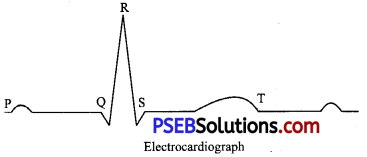
- The T-wave represents the return of the ventricles from excited to normal state (repolarisation).
- The end of the T-wave marks the end of systole.
- Obviously, by counting the number of QRS complexes that occur in a given time period, one can determine the heartbeat rate of an individual.
- Since the ECGs obtained from different individuals have roughly the same shape for a given lead configuration, any deviation from this shape indicates a possible abnormality or disease.
- Hence, it is of a great clinical significance.